


Research_AdobeStock_©-Gorodenkoff_201606406_2000px_191205

Research

Medical Omics

251121A_BIH_Logo_RGB_BIH_Logo_Standard_BlauKorall
ChariteUMB-Logo_RGB_600px_191023

Medical Omics Overview

Berlin

The Medical Omics lab aims to improve healthcare and basic research by providing artificial intelligence-driven tools for the analysis of large biomedical datasets. For comprehensive insights in disease entities, we aim to include multiple layers of data, such as single-cell RNA sequencing, methylomics data and medical imaging. In close collaborations with clinicians at the Charité, we develop clinical decision support systems for cancer treatment as well as solutions for routine clinical tasks such as automated annotation of radiological images.

Topics

Oncology

Our research in oncology is centered on developing computational methods that improve the molecular diagnosis and imaging-based analysis of cancer, with a particular focus on brain tumors.

One major area of our work is the development of machine learning methods for DNA methylation-based tumor classification. DNA methylation patterns provide a molecular fingerprint that can distinguish between different tumor types with high accuracy and have become an important component of modern cancer diagnostics. We develop algorithms that make these approaches more flexible, robust, and widely applicable across different sequencing technologies. Our methods enable accurate classification even from sparse or incomplete methylation data, allowing reliable diagnosis across more than 170 cancer types while supporting rapid and cost-effective clinical workflows leveraging modern sequencing technologies such as Nanopore sequencing for ultra-fast diagnostics (Yuan et al., Nat Cancer 2025).

In addition to molecular diagnostics, we develop deep learning methods for the automated analysis of medical images. This includes segmentation of gliomas and organs-at-risk in magnetic resonance imaging (MRI), enabling precise identification of tumors and their different tissue components as well as areas particularly sensitive to radiation therapy. Automated image analysis reduces the need for time-consuming manual annotations while providing accurate and reproducible tumor delineation, supporting treatment planning and disease monitoring (Jabareen & Lukassen, MICCAI BrainLes 2021).

Together, our research combines artificial intelligence, genomics, and medical imaging to develop computational tools that contribute to more precise, faster, and more scalable cancer diagnosis and personalized patient care. 

Cardiology

Our research in cardiology aims to develop artificial intelligence methods for the analysis of cardiovascular data, with a particular focus on electrocardiograms (ECGs) and echocardiography. By combining advances in machine learning with close collaboration with clinical and industry partners, we aim to improve the accuracy, reliability, and efficiency of computer-assisted cardiac diagnostics (FACE project).

A major focus of our work is the development of deep learning models for automated ECG interpretation. We investigate how neural network architectures can be specifically optimized for physiological signals rather than directly adopting designs from computer vision. By systematically studying how model architecture and size influence performance, we derive ECG-specific design principles that enable more accurate and computationally efficient classification of cardiac abnormalities (Jabareen et al., ESC Digital Health 2025).

To ensure that these models perform reliably in real-world clinical settings, we study their robustness across different ECG recording modalities, including resting, telemedical, and long-term ECGs. Our research addresses the challenges arising from variations in recording conditions and develops methods that recognize domain shifts and quantify predictive uncertainty. This allows models to indicate when their predictions may be less reliable, increasing their safety and trustworthiness in clinical practice (Zillekens et al., ESC Digital Health 2025).

We also investigate machine learning methods tailored to highly imbalanced clinical datasets, where detecting a small number of clinically relevant cases is particularly important. By designing novel optimization strategies that directly target clinically meaningful evaluation metrics, we improve the identification of high-risk patients while maintaining robust model performance (Herzler et al., CinC 2025).

Beyond ECG analysis, we explore the application of multimodal foundation models to cardiac ultrasound imaging. Our work evaluates the ability of modern vision-language models to interpret the fine-grained temporal dynamics of echocardiography, revealing important limitations of current general-purpose AI systems and highlighting the need for domain-specific approaches for cardiac video analysis (Liu et al., MIDL 2026).

These methodological advances are integrated into practical clinical workflows through projects that combine edge computing and cloud-based AI. By distributing data processing between local devices and centralized infrastructure, our systems enable rapid ECG analysis, continuous model improvement, and seamless support for clinicians, ultimately contributing to more efficient and reliable cardiovascular care.

Methods development

Our group develops novel deep learning methods that address fundamental challenges in biomedical data analysis. Classical machine learning algorithms are often ill-suited to specific properties of medical data, such as absolute distance and position measure in CT and MRI imaging. Thus, we aim to adapt standard techniques to these properties, and re-develop new ones where needed. This section outlines our work on more fundamental concepts of deep learning for biomedical data.

A central area of our work is representation learning, where we develop models that automatically extract meaningful information from complex datasets without requiring extensive manual annotation. This includes self-supervised learning methods for medical imaging that exploit the inherent spatial organization of anatomical structures, enabling the learning of informative image representations from large collections of unlabeled data (Jabareen et al., arXiv 2024).

We also investigate new deep learning architectures for biological data, particularly single-cell sequencing. Our methods combine probabilistic deep learning with interpretable latent representations to identify biologically relevant gene sets, characterize cellular heterogeneity, and quantify the confidence of inferred features. By integrating multiple data modalities and leveraging ensemble learning, we improve the robustness and reproducibility of computational analyses while reducing the need for extensive manual feature engineering (Lukassen, Ten et al., Nat Mach Intell 2020 and Ten et al., Front Cell Dev Biol 2023).

Another focus is the development of methodological advances for modern neural network architectures. We study how fundamental design choices, such as positional encoding in Transformer models, influence learning in medical imaging and develop new architectures that explicitly account for the spatial and anisotropic properties of biomedical data. These methodological innovations improve both model performance and interpretability across a wide range of imaging modalities (Jabareen et al., MICCAI ShapeMI 2025).

Together, this research provides the methodological foundation for many of our application-oriented projects in oncology, cardiology, and computational biology, while contributing broadly applicable machine learning techniques to the biomedical AI community.
Sören_Lukassenjpg

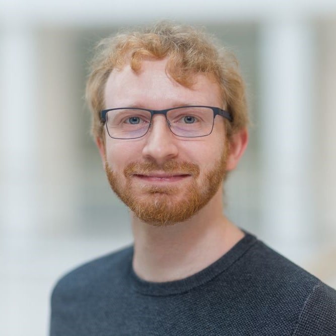
Sören Lukassen studied Molecular Medicine and received a PhD in human genetics from the FAU Erlangen-Nürnberg, where he specialized in (single-cell) sequenzing data analysis and deep learning. He joined Christian Conrads lab in Heidelberg in 2018 and moved to Berlin the same year. Since 2021, he leads the junior research group Medical Omics, which focuses on integrating data from multiple sources for diagnosis and treatment of diseases. 

Dr. Sören Lukassen

Group leader Medical Omics
BIH@Charité - Center of Digital Health
Luisenstr. 65
10117 Berlin
 

Research Group

Florian Herzler
research assistant
florian.herzler@bih-charite.de
Dingming Liu
PhD Student
dingming.liu@charite.de

Projects

FACE


Publications

Jabareen N., and  Lukassen S.*, (2022). Segmenting brain tumors in multi-modal MRIscans using a 3D SegNet architecture. MiCCAI BrainLes, https://doi.org/10.1007/978-3-031-08999-2_32,   DOWNLOAD

Trump S.*, Lukassen S.*, Anker M.S.*, Chua R.L.*, Liebig J.*, Thürmann L.*, Corman V.M.*, Binder M., Loske J., Klasa C., Krieger T., Hennig B.P., Messingschlager M., Pott F., Kazmierski J., Twardziok S., Albrecht J.P., Eils J., Hadzibegovic S., Lena A., Heidecker B., Bürgel T., Steinfeldt J., Goffinet C., Kurth F., Witzenrath M., Volker M.T., Muller S.D., Liebert U.G., Ishaque N., Kaderali L., Sander L.E., Drosten C., Laudi S., Eils R., Conrad C., Landmesser U., Lehmann I. (2021) Hypertension delays viral clearance and exacerbates airway hyperinflammation in patients with COVID-19. Nature Biotechnology doi: 10.1038/s41587-020-00796-1

Chua, R.L.*, Lukassen, S.*,Trump, S.*, Hennig, B.P.*, Wendisch, D.*, Pott, F., Debnath, O., Thürmann, L., Kurth, F., Völker, M.T., Kazmierski, J., Timmermann, B., Twardziok, S., Schneider, S., Machleidt, F., Müller-Redetzky, H., Maier, M., Krannich, A., Schmidt, S., Balzer, F., Liebig, J., Loske, J., Suttorp, N., Eils, J., Ishaque, N., Liebert, U.G., von Kalle, C., Hocke, A., Witzenrath, M., Goffinet, C., Drosten, C., Laudi, S.§,Lehmann, I., Conrad, C., Sander, L.-E. & Eils, R. (2020). COVID-19 severity correlates with airway epithelium-immune cell interactions identified by single-cell analysis. Nature Biotechnology doi: 10.1038/s41587-020-0602-4

Lukassen, S.*, Chua, R. L.*, Trefzer, T.*, Kahn, N.C.*, Schneider, M.A.*, Muley, T., Winter, H., Meister, M., Veith, C., Boots, A.W., Hennig, B.P., Kreuter, M., Conrad, C., & Eils, R. (2020). SARS-CoV-2 receptor ACE2 and TMPRSS2 are primarily expressed in bronchial transient secretory cells. EMBO Journal doi: 10.15252/embj.20105114

Lukassen S.*, Ten F.W.*, Adam L., Eils R., Conrad C. (2020). Gene set inference from single-cell sequencing data using a hybrid of matrix factorization and variational autoencoders. Nature Machine Intelligence doi: 10.1038/s42256-020-00269-9


*These authors contributed equally


Latest News

de.KCD Industry Forum

As part of the de.KCD/de.NBI network the Center of Digital Health organizes this year’s de.KCD Industry Forum on June 18, 2026 at the Einstein Center Digital Future in Berlin. de.KCD creates a forum
SBHD 2026

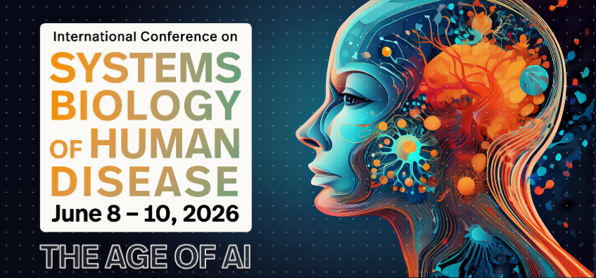
Our colleagues from Northestern University in Boston are organising the 18th INTERNATIONAL CONFERENCE ON SYSTEMS BIOLOGY OF HUMAN DISEASE – SBHD 2026 in Boston from June 08-10. This years Conference
Development of the European Cancer Information Portal Launched

The Berlin Institute of Health at Charité (BIH) coordinates the pan-European research initiative "EU-CiP" (European Cancer Information Portal). Supported by 12 million euros from the EU’s Horizon
SBHD 2025

The Digital Health Center once again organises the INTERNATIONAL CONFERENCE ON SYSTEMS BIOLOGY OF HUMAN DISEASE – SBHD 2025 in Berlin from June 16-18. Don’t miss this opportunity to participate in a