


Christian Conrad graduated in Biology at the University Freiburg and received his PhD in Bioinformatics at the University Heidelberg. In 2018, he moved to the Charité/BIH where he was appointed as Professor for Intelligent Imaging.
Prof. Dr. Christian Conrad
Group leader Intelligent Imaging
Post Address:
Charité - Campus Charité Mitte | Charitéplatz 1 | 10117 Berlin | Germany
Visiting Address:
Rahel Hirsch Center | Luisenstraße 65 | 10117 Berlin
Level 02, Part B, room 217
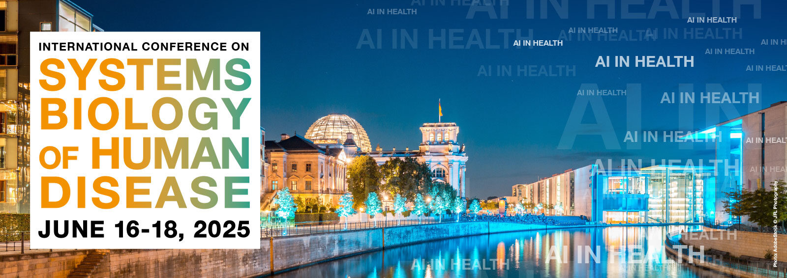
The Digital Health Center once again organises the INTERNATIONAL CONFERENCE ON SYSTEMS BIOLOGY OF HUMAN DISEASE – SBHD 2025 in Berlin from June 16-18. Don’t miss this opportunity to participate in a
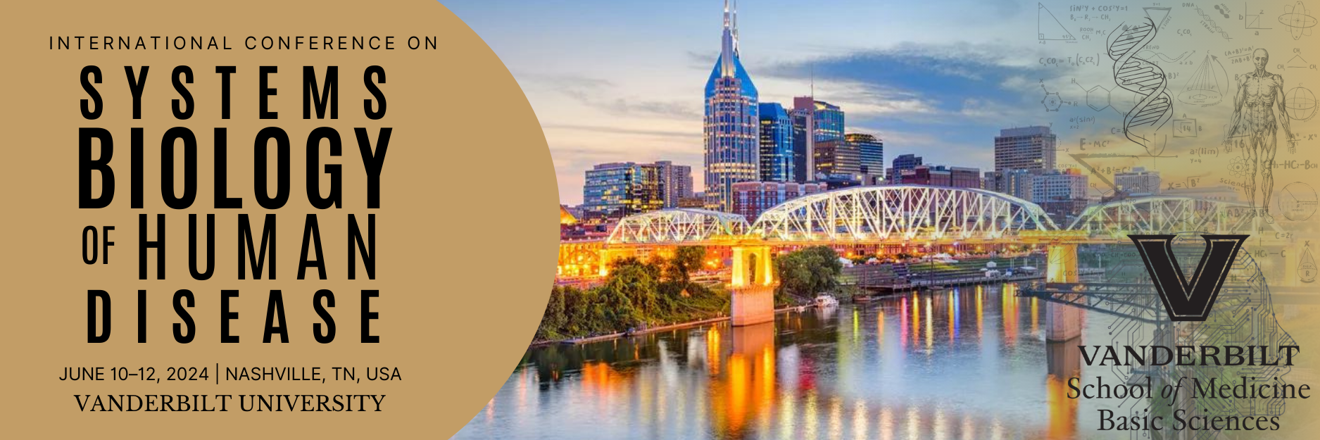
Our colleagues at Vanderbilt University organise the 16th INTERNATIONAL CONFERENCE ON SYSTEMS BIOLOGY OF HUMAN DISEASE – SBHD 2024 this year from June 10-12. Don’t miss the opportunity to participate

Am 11.10.2023 wurde Prof. Roland Eils im Tagesspiegel als einer der 100 wichtigsten Köpfe der Hauptstadt-Wissenschaft gewürdigt. So schreibt der Tagesspiegel: "Um Big Data dreht sich alles in der
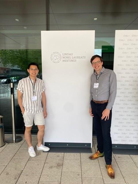
The Lindau Nobel Laureate Meetings are annual conferences where some of the brightest minds in science converge to exchange knowledge, foster collaboration, and inspire the next generation of